
AI assists live brain surgery for the first time, and the patient can see clearly again
Surgeons in London removed a pituitary tumor from a 48-year-old with help from an AI that analyzed the live camera feed and color-coded nerves and blood vessels. The hospital calls it the world's first neurosurgery with real-time AI assistance. It doesn't yet prove that such operations become safer.
What's going on
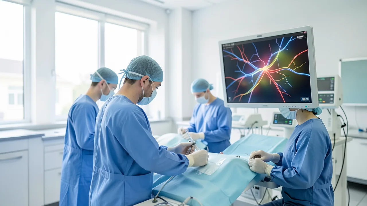
In May 2026, neurosurgeons at the National Hospital for Neurology and Neurosurgery (NHNN) in London, part of University College London Hospitals (UCLH), removed a brain tumor with the help of real-time AI. According to UCL and UCLH, it's the world's first neurosurgery in which an AI assisted live during the operation. Details were only released after the patient had recovered.
The patient is Rhys Hibbert, 48, from Bedfordshire, England. In December 2024, he collapsed while out on a walk and had his first epileptic seizure. Scans revealed a tumor of about 11 millimeters on his pituitary gland, a marble-sized organ at the base of the skull that controls almost all of the body's key hormones. According to Techtimes, it was a pituitary adenoma, a benign tumor. At this size it's called a macroadenoma.
The tumor was pressing on his optic nerves. Hibbert's field of vision narrowed until he could no longer see the lower part of it. He also had a severe hormonal disorder, fatigue, dizziness and balance problems. Because he kept stumbling, he walked with canes. Doctors first treated him without surgery, but when his symptoms worsened over more than a year and, according to the hospital, he was at risk of going blind, surgery became unavoidable. He agreed to be the first patient operated on with the AI assistant.
Why this surgery is so tricky
The pituitary gland is usually operated on through the nose. An endoscope, a thin instrument with a camera on its tip, is pushed through the nostril and sphenoid sinus up to the base of the skull. There, the pituitary sits in a bony hollow called the sella turcica. Right next to it run the carotid arteries, the optic chiasm and venous sinuses. According to Techtimes, the margin for error is about one millimeter. A small navigation mistake can mean blindness, a stroke or death.
Until now, surgeons have mostly relied on navigation systems that match MRI scans taken before surgery to the patient's position. The problem: during surgery, tissue shifts, it bleeds, cerebrospinal fluid drains and tumor tissue gets removed. According to Techtimes, this displacement, known as "brain shift," can amount to several millimeters. That's enough to make the pre-op map useless exactly when it's needed most.
How the AI works
The system was developed at UCL's Hawkes Institute, with Dr. Sophia Bano, Associate Professor of Robotics and AI, as technical lead. Instead of analyzing old scans, it analyzes the endoscope's live video. It sees exactly what the surgeon sees, just analyzed frame by frame.
According to Techtimes, it's a convolutional neural network (CNN), a type of neural network that's especially good at recognizing patterns in images. It was trained on a large collection of videos from previous pituitary operations. Neurosurgeons annotated individual frames: where the sella is, where important vessels run, where the tumor ends. The model then performs semantic segmentation, assigning every single pixel to an anatomical structure, and displays the result color-coded on a second monitor next to the main screen. The system is reportedly running on Nvidia's Clara IGX platform, hardware built specifically for fast AI computation in medical devices.
According to The Next Web, an earlier version was presented publicly in 2023, trained at the time on more than 200 surgical recordings. Back then, the researchers expected AI assistance in brain surgery within about two years. UCLH says the system gained as much surgical experience in around ten months as a trainee surgeon would in about a decade. Sophia Bano says:
"By learning from hundreds of surgical videos, it has seen a range of surgical examples that would take a surgeon many years."
Importantly, the AI didn't control any instruments or make a single decision. The operation was performed by Prof. Hani Marcus of the UCL Queen Square Institute of Neurology and Danyal Khan, a resident and PhD student who leads the research program. Marcus describes the system as a second pair of expert eyes that never gets tired.
What the data so far shows
The team has published earlier work. A November 2024 study in npj Digital Medicine tested an earlier model offline, on still images rather than in the operating room. Participants had to outline the sella on endoscope images, once with and once without AI help. Performance was measured with the DICE score, which captures how closely a drawn area matches the correct one.
| Group | without AI | with AI |
|---|---|---|
| All participants | 70.7% | 77.5% |
| Medical students | 66.2% | 78.9% |
| AI alone (test dataset) | – | 79.1% |
According to The Next Web, this model was based on 640 images from 64 surgical videos. Notably, experienced surgeons benefited less than beginners. Techtimes concludes that the biggest payoff may not be at top centers but at hospitals that perform these procedures less often.
A survey of surgeons in the journal Surgical Endoscopy (June 2026) found, according to Techtimes, that 86.8 percent consider AI in the operating room helpful. Anatomy recognition got the highest usefulness score at 4.57 out of 5. Yet this kind of technology is still rarely used in everyday practice.
The outcome, and what it doesn't prove
According to UCL, the tumor was successfully removed and the patient's vision preserved. Hibbert told the Guardian: "When I woke up, I could see everything in the room clearly." Within a week, he was walking without glasses or canes, and he's now back at work as a customer service manager. "It feels like I've got a 360-degree panoramic view," he says.
Gadgetreview rightly points out, though, that these are the patient's own accounts, not independently measured trial results. And one successful operation says nothing about whether the AI reduces complications or beats conventional surgery. That takes larger studies with control groups. The Next Web notes that the system has no public name, no accuracy or outcome figures from the operation have been released, and the trial's name, size, design and regulatory status are all unknown. The system isn't approved for routine use outside the trial.
Techtimes lists known weaknesses of such systems: performance drops when blood, smoke or lots of instruments obscure the view, rare anatomical variants are underrepresented in training data, and a system trained at one hospital doesn't always work as well elsewhere. It's also unclear what happens when the AI misidentifies a structure, and who's responsible when a surgeon disagrees with what it shows. Displaying the guidance on a separate screen also forces surgeons to split their attention between two monitors, something the team wants to improve. The next version is meant to track instruments and their contact with tissue in real time as well.
The study was funded by the National Institute for Health and Care Research (NIHR) and, according to Techtimes, also by Google, with support from organizations including the Royal College of Surgeons and Wellcome. Prof. Mike Lewis of the NIHR called it "groundbreaking surgery."
Our take
This case shows that AI image analysis can actually make the jump from the lab to the operating room: in real time, on the live feed, as an aid rather than a replacement for the surgeon. That's a genuine feasibility milestone. The most exciting idea is putting the experience of hundreds of operations at the fingertips of less experienced surgeons. But this was a single procedure, and hard data from the operation is missing. Whether the technology really makes surgery safer will have to be shown by larger, independently evaluated studies, which the UCL team says it's working on. The NHNN was founded in 1859 as the world's first dedicated neurosurgical hospital, and Hibbert finds it fitting that this first happened there too.
Sources
- Merkliste: gadgetreview.com gadgetreview.com
- techtimes.com techtimes.com
- techspot.com techspot.com
- thenextweb.com thenextweb.com
Ask the AI assistant
Questions about this article, installation or background? Our assistant knows this page and runs on our own machine, not in someone else's cloud.
The Assistant is an AI (Gemma 4) and can make mistakes – please check important details against the source. Questions are used only to generate an answer and are deleted after one hour. Please don't enter any personal data.


Comments
What do you think? Already tried it? Write it here, no sign-up needed.
Comments are public. Links aren't allowed, and we delete abusive posts. You can delete your own comment at any time in this browser. Learn more in the Privacy Policy.